Showing posts with label chapter 6. Show all posts

Wednesday, January 20, 2016

Biology: Chapter 6: Nucleic acids and protein synthesis: Protein synthesis

Biology: Chapter 6: Nucleic acids and protein synthesis: Protein synthesis

  • Protein synthesis: Process in which the bases on the DNA molecule is used to code for the sequence of amino acids in a polypeptide.
  • Sequence of amino acids determine the 3-D folding of the protein and therefore it's function.

Transcription

  • A section of the DNA (gene) has the instructions for creating a mRNA (messenger RNA) molecule.
  • Promotor: Signals the start of a gene.
  • Terminator/ Stop sequence: Signals the end of the gene.
  • RNA polymerase: Binds to the promotor and when the DNA uwinds initiates RNA synthesis.
  • Sense strand: DNA strand with the same code as the mRNA.
  • RNA polymerase starts RNA synthesis as the DNA unwinds at the promotor. 
  • Moves down the DNA strand in a 5' to 3' direction, until it reaches the base code for the stop sequence at the end of the gene, where the newly formed RNA transcript peels off the template strand.
  • DNA rewinds back into the double helix after the RNA synthesis.
  • The mRNA breaks away from the DNA and leaves the nucleus through the nuclear pores into the cytoplasm.
  • mRNA (messenger RNA): RNA transcript created in translation.
  • Template strand: DNA strand that the mRNA uses as a template for RNA synthesis - the antisense strand.
Simple diagram of transcription.

Translation

  • Process of building chain of amino acids following code on mRNA molecule made in transcription.
  • mRNA leaves nucleus through nuclear pores into the cytoplasm.
  • Ribosome (made of rRNA - ribosomal RNA) clamps around the mRNA.
  • Ribosomes: Responsible for protein synthesis.
    Have 2 units - large subunit and small subunit, which clamp around the mRNA.
    The large subunit has 3 sites - A siteP site and E (exit) site.
    These are for tRNA (transfer RNA) to come in and bond to it's complementary codon.
    Two tRNA molecules can be in a ribosome at one time.
    Ribosome.
  • Codon: Groups of 3 bases which code for an amino acid
  • Anti-codon: 3 bases on a tRNA. Forms a bond with it's complementary codon on the mRNA strand in the ribosome.
  • tRNA (transfer RNA): Made from a single strand of RNA, twisted to form a 3 leaf clover shape. Has a anti-codon (base triplet) on one end, and amino acid attachment site on the other.
    tRNA molecule.
  • The tRNA bonds to an amino acid specific to it's anticodon and carry it to the ribosome, where it forms hydrogen bonds with it's complementary codon on the mRNA strand.
  • 2 tRNA molecules are in the ribosome at one time. The ribosome cuts the bonds of the amino acid and the tRNA in the P site, and the amino acid forms a peptide bond with the amino acid on the tRNA in the A site, forming a amino acid chain.
  • The ribosome then moves down the mRNA chain, in a 5' to 3' direction. 
  • The tRNA that was previously in the P site moves to the E site, and is released from the ribosome. 
  • The tRNA in A is now in P, and a new tRNA enters the A site
  • The ribosome again cuts the bond between the amino acids and tRNA in the P site, which again form a peptide bond with the amino acid on the tRNA in the A site
  • The process continues, forming a polypeptide chain.
    Ribosome in translation.
  • start codon signals the start of translation in the ribosome - usually AUG
  • When the stop codon is reached on the mRNA strand - usually UAG, UAA or UGA, the ribosome accepts a release factor, which allows the ribosome to break the bond between the last tRNA and it's now polypeptide chainreleasing the polypeptide chain, which will fold into secondary, tertiary and quaternary structures according to it's amino acid sequence.
  • The ribosome and tRNA seperate from the mRNA chain.
  • There are 20 different types of amino acids present in the cytoplasm.
  • Many ribosomes work on the mRNA strand at one time, so multipe copies of the polypeptide can be made efficiently.
    Translation diagram.

Mutations

  • Changes in the genetic material.
  • Change of bases in the code, which can lead to a new gene (alleles).
  • Alleles: Different types of the same gene (Blue or brown eyes).
Point mutations - Changes in just one base pair
  • Substitution: Replacement of one nucleotide and it's complementary base in the DNA strand. Leads to alteration in amino acid sequence, which can hinder protein function eg. Sickle cell anemia - glutamic acid replaced with valine.
  • Insertion/Deletion: nucleotide pair inserted or removed in the DNA strand. Can result in an extra or missing amino acid. May alter reading frame (codon groups) - Frameshift mutation, which creates a protein that is almost certainly unable to function.
DNA mutations.

Monday, January 18, 2016

Biology: Chapter 6: DNA and Nucleic acids: DNA replication

Biology: Chapter 6: DNA and Nucleic acids: DNA replication

  • In 1953, James and Watson Crick used the results of work by Rosalin Franklin to develop a model of the structure of DNA.
  • Semi-conservative replication: Method of copying where half of the original molecules are kept (conserved) in the new molecules.
    Simple diagram of semi-conservative replication in DNA.
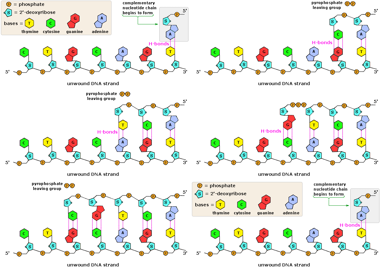
  • Enzyme Helicase unzips parental double helix, separating the two strands.
  • Replication fork: Where the DNA strands are separating.
    Replication fork.
  • Single-strand binding proteins: Keep the two parental strands separated by temporarily hydrogen bonding to the bases.
  • Primase: Adds RNA primer to DNA strand
  • RNA primer: Initiate DNA synthesis by DNA polymerase
  • DNA polymerase: Builds a new strand by assembling the nucleotides to the parental strand. Nucleotide bases form the specific bonds with its complementary base on the parental strand, therefore it is almost impossible to make a mistake. Only works in a 5' to 3' direction.
  • Since DNA polymerase can only work from a 5' to 3' direction, one strand can be built continuously as the replication fork progresses. 
  • Leading strand: Strand that is built continuously from from 5' to 3'.
  • However, one strand will have the replication fork progressing towards 5'. This strand will grow in the overall direction of 5' to 3' by the addition of short fragments as the replication fork progresses. 
  • Lagging strand: 3' to 5' strand that needs to be built in fragments.
  • Okazaki fragments: Fragments used to build the 3' to 5' strand.
  • Another DNA polymerase replaces RNA primer with DNA.
  • DNA ligase: Joins okazaki fragments together to growing strand.
DNA replication diagram.

Proofreading DNA
  • DNA polymerase proofreads each nucleotide and removes incorrectly paired nucleotides.
Repairing DNA
  • DNA molecules that are exposed to harmful chemical and physical agents eg. Tobacco, UV rays can change nucleotides, which can affect encoded genetic information. However, the DNA has some ways of fixing it.
Nucleotide excision repair:
  • Nuclease enzyme: Cuts damaged DNA strand at it's 2 points.
  • Repair synthesis by DNA polymerase fills gap
  • DNA ligase: Seals the phosphate sugar backbone together.

Friday, January 15, 2016

Biology: Chapter 6: Nucleic acids and protein synthesis: DNA and RNA structure

Biology: Chapter 6: Nucleic acids and protein synthesis: DNA and RNA structure

Nucleotides aka Nucleic acids

  • Smaller molecules that DNA and RNA are made up of.
  • Polynucleotides: Long chains of nucleotides eg. DNA and RNA.
  • Composed of 3 components:
    -Pentose sugar
    -Nitrogen-containing base
    -Phosphate group
Structure of nucleotide.
  • Pentose sugar: Sugar with 5 carbons. Can be either Ribose (in RNA) or Deoxyribose (in DNA.
  • Deoxyribose: Missing an oxygen on it's second carbon primer.
Nitrogen-containing bases aka Nitrogenous bases
  • Purine bases: Double ring structure - Adenine, Guanine.
  • Pyrimidine bases: Single ring structure - Thymine, Cytosine, Uracil.
  • DNA contains the 4 bases A-T and C-G, but never Uracil.
  • RNA contains the 4 bases A-U and C-G, but no Thymine.
Structural formulae of the nitrogenous bases.
ATP: Similar structure to a nucleotide. Adenosine (Ribose sugar with the base Adenine) can be combined  to 1,2 or 3 phosphates to make Adenosine Monophosphate (AMP), Adenosine Diphosphate (ADP) or Adenosine Triphosphate (ATP).

Polynucleotides
  • Polynucleotide: Many nucleotides linked together in a chain
  • Formed during interphase of the cell cycle.
  • Sugar-phosphate backbone: Alternating sugars and phosphates of the nucleotides linked together with the bases projecting sideways.
  • Phosphodiester bonds: Covalent sugar-phosphate bonds.
  • Phosphodiester bonds link the Carbon 5 of one sugar to the Carbon 3 of the next.
  • The polynucleotide is said to have 3' and 5' ends: the end where the Carbon 3 has nothing to link is called the 3' end, while the end where the Carbon 5 has nothing to link to is called the 5' end.
  • Bases can be purines or pyrimidines: Purines only link with pyrimidines, but it is even more precise.
  • In a DNA molecule, there is only enough room between the two sugar-phosphate backbones for 1 purine and 1 pyrimidine molecule, so a pyrimidine can only bond with a purine, since purines are larger.
  • Adenine and Thymine form double hydrogen bonds, whereas Guanine and Cytosine form a triple hydrogen bond, therefore only pairs possible are A-T and C-G.
  • Complementary base pairing: The precise pairing of bases due to hydrogen bond and structure size (purine/pyrimidine) : Adenine and Thymine (A-T), and Cytosine and Guanine (C-G).
Animation of DNA replication and the DNA strand.

  • Double helix: Two strands wrapping around each other - the 3D shape DNA molecules form.
  • Hydrogen bonds linking the bases easily broken, which happens during DNA replication and protein synthesis. 
  • RNA molecules are only single stranded.