Monday, November 30, 2015

Biology: Chapter 4: Cell surface membrane: Movement in and out of cells

Biology: Chapter 4: Cell surface membrane: Movement in and out of cells

Five basic mechanisms by which exchange is achieved:
  1. Diffusion
  2. Faciliated diffusion
  3. Osmosis
  4. Active transport
  5. Bulk transport

Diffusion

  • Net movement of particles from a region of high concentration to a region of lower concentration through random movement of it's molecules.
  • Move down a concentration gradient
  • Random movement caused by natural kinetic energy
  • To reach equilibrium
Factors that affect diffusion rate across a membrane:
  • Steepness of concentration gradient: The greater the difference in concentration, the faster the rate of diffusion.
    More molecules will be moving from one side to another
  • Temperature: The higher the the temperature, the faster the rate of diffusion
    Higher temperature means more kinetic energy - molecules move faster
  • Surface area: The greater the surface area, the faster the rate of diffusion
    -The greater the surface area, the more molecules can cross it at any one moment.
    -Surface area can be increased by folding 
    -The larger the cell, the smaller it's surface area is in relation to it's volume -volume increases more rapidly than surface area as size increases.
    -Therefore there is a limit on the size of cells, since they rely on diffusion for internal transport.
    -Time it takes for a molecule to reach it's destination by diffusion increases rapidly with distance traveled.
    -Most cells are only 50 micrometers in diameter.
  • Nature of molecules or ions: Small, non-polar molecules diffuse faster through the membrane (eg. oxygen and carbon dioxide diffuse directly through phospholipid bi-layer).
    -Non-polar molecules are not repelled by the hydrophobic interior of the phospholipid bi-layer.
    -Large molecules require more energy to move, therefore diffuse more slowly.
    -Water molecules, despite being polar, diffuse directly through the bi-layer because they are small enough to not be repelled by hydrophobic tails.

Facilitated diffusion

  • Diffusion of large polar molecules and ions made possible by transport proteins.
  • Two types of transport proteins: channel and carrier proteins.
  • Each is highly specific and only lets one type of molecule/ion to pass through it.
Channel proteins
  • Water-filled pores
  • Fixed shape
  • Allow charged substances usually ions, to diffuse through membrane.
  • Usually 'gated' - Part of the protein molecule on the inside surface of the membrane can move to close or open the pore, like a gate.
  • This allows control of ion exchange.
  • eg. Nerve cell surface membrane channel proteins; one type allows entry of sodium ions for production of an action potential, while another allows the exit of potassium ions during recovery phase for repolarisation.
  • Some channels occur in a single protein while others are formed by several proteins combined.
Carrier proteins
  • Flip between two shapes 
  • The binding site alternately opens to one side of the membrane, then the other.
  • The molecules will move down the concentration gradient across the membrane like in normal diffusion.
  • Rate of diffusion depends on how many channel and carrier proteins the membrane has.
  • In the case of carrier proteins, the rate of diffusion also depends on whether they are open or not.
  • Cystic fibrosis is caused by a deficit in a channel protein that allows chloride ions to move out of the cells lining the lungs.

Osmosis

  • Special type of diffusion involving only water molecules.
  • Movement of water molecules by diffusion from a dilute solution to a concentrated solution through a partially permeable membrane.
  • Remember: Solute + Solvent = Solution
  • Two solutions separated by a partially permeable membrane - only allows certain molecules through.
Representation of the plasma membrane   A                                                         B
  • In solution A, there are more sugar molecules on the left than the right - the left is more concentrated
  • The partially permeable membrane only allows water molecules through.
  • The water molecules diffuse through the membrane from the right to the left, down a concentration gradient due to random movements as to reach equilibrium
  • In solution B, the left contains more water molecules, so it is more dilute, and the right contains less water, becoming more concentrated.
Water potential
  • Tendency of water to move out of a solution.
  • Depends on two factors:
    -How much water the solution contains in relation to solutes (concentration)
    -How much pressure is being applied
  • Water potential always moves down a water potential gradient (region of high water potential to a region to low water potential) until water potential is same throughout the system - equilibrium.
  • A solution containing a lot of water (dilute) has a higher water potential than a solution containing a little water (concentrated).

  • Increasing pressure on a solution also increases water potential - increases the tendency for water to move out of it.
  • Water potential of pure water at atmospheric pressure is 0.
  • Therefore a solution (water with solute/solutes dissolved in it) must have a negative water potential - less than 0.
Solute potential and pressure potential
  • Solute potential: Extent to which solute molecules decrease the water potential of the solution.
  • Solute potential is also 0 for pure water, and a negative value for a solution
  • Adding more solute to a solution decreases it's water potential.
  • Pressure potential: Contribution of pressure to the water potential of a solution.
  • Increasing pressure increases water potential.
Osmosis in animal cells
  • If the water potential surrounding the cell is too high - cell swells and bursts (lysis)
  • If it is too low - cell shrinks
  • Essential to maintain a constant water potential in the bodies of animals 
Osmosis in a blood cell

  • Hypotonic: Water potential surrounding cells too high
  • Isotonic: Normal water potential surrounding cells
  • Hypertonic: Water potential surrounding cells too low 
Osmosis in plant cells
  • Unlike animal cells, plants have a cell wall.
  • Cell walls are freely permeable.
  • When water potential is higher outside than inside the cell, water will enter the cell. 
  • However, as water enters plant cells the cell wall will push back against the expanding protoplast (living part of the cell) and pressure builds up.
  • This is the pressure potential and it increases the water potential until the water potential inside and outside the cell are equal - equilibrium is reached
  • The cell wall is so inelastic that it takes very little water to enter the cell to achieve this.
  • Cell wall prevents cell from bursting.
  • A plant cell is fully turgid when it is fully inflated with water.
  • Water potential = Solute potential + Pressure potential 
Osmosis in a plant cell
  • Hypotonic: Fully turgid
  • Isotonic: No net movement of water
  • Hypertonic: Water leaves cell and protoplast gradually shrinks until it exerts no pressure on the cell wall - pressure potential 0.
    As the protoplast continues shrinking it
    begins to pull away from the cell wall.
    This is called
    plasmolysis.
    When the protoplast has completely shrunken away from the cell wall, it is said to be
    fully plasmolysed. When the pressure potential has just reached 0 and plasmolysis is about to occur is called incipient plasmolysis

Active transport 
  • Movement of molecules or ions through carrier proteins across a cell membrane against their concentration gradient using energy from ATP.
  • ATP (produced during cell respiration) is used to make the carrier proteins change its shape, transferring molecules or ions across the membrane in the process.
  • Can occur either into or out of the cell.
  • Example: Sodium potassium (Na+ - K+) pump
  • Found in cell surface membranes of all animal cells
  • Run almost all the time, on average use 30% of a cells energy.
  • For each ATP molecule used it pumps 3 sodium ions out of the cell while at the same time allowing 2 potassium ions into the cell.
  • Net result is the inside of the cell becomes more negative than outside - potential difference (p.d) is created across membrane.
  • The pump has a receptor for ATP on it's inner surface. The receptor acts as an ATPase enzyme in bringing about the hydrolysis of ATP to ADP (adenosine diphosphate) and phosphate to release energy.
Sodium potassium pump

  • Active transport is also important in re-absorption of ions and useful molecules in the kidneys after filtration and absorption of some products of digestion from the gut. In plants, active transport is used inorganic ions from soil to the root hairs and load sugars into the phloem tissues.

Bulk transport

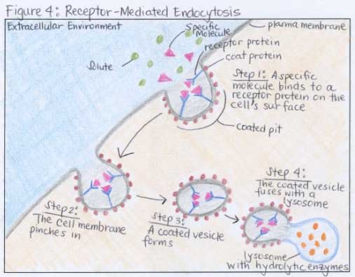
  • Bulk transport of large quantities of materials into (endocytosis) and out of (exocytosis) of cells.
  • Endocytosis: Engulfing of a material by the cell surface membrane to form a small sac (endocytic vacuole). Two forms:
         -
    Phagocytosis (cell eating): Bulk uptake of solid material. Cells specialising in this are called phagocytes, and the vacuoles are called phagocytic vacuoles. Example: White blood cells engulfing bacteria. Phagocytic vacuoles fuse with lysosomes, which contain digestive enzymes.
         -
    Pinocytosis (cell drinking): Bulk uptake of liquid. Vacuoles formed are often really small, in which the process is called micropinocytosis
  • Protein receptors on the outer cell surface membrane detect the molecules that need to be transported and binds to them.
Endocytosis

  • Exocytosis: Reverse of endocytosis - materials are removed from cells. Usually involves golgi body. Example: In secretion of enzymes, secretory vesicles from the golgi body carry the enzymes to the cell surface to release their contents
Exocytosis

 Simplified exocytosis and endocytosis


Thursday, November 26, 2015

Biology: Chapter 4: Cell surface membrane: Cell signalling

Biology: Chapter 4: Cell surface membrane: Cell signalling

  • Getting a message from one place to another.
  • Complex range of signalling pathways which coordinate activities of the cell so they respond appropriately to the environment, even if the cells are large distances apart in the body.
  • Signalling pathway includes:
  1. Receptor receiving a stimulus or signal
  2. Transduction: Converting the signal to a message that is readable 
  3. Transmission of message/signal to effector (target)
  4. The effector making an appropriate response

  • Distances can be short (diffusion within a cell) or long (transport through blood)
    -Endocrine signalling: Signalling over large distances, often through circulatory system
    -Paracrine signalling: Signalling occurring between cells close together either through extracellular fluid or directly between cells
    -Autocrine signalling: Cell stimulates response within itself by releasing signals for it's own receptors

  • Many components and different mechanisms along the route
  • Signalling includes both electrical (nerves) and chemical (hormones).
  • Stimuli can be from inside (hormones) as well as outside (light).
  • The cell surface membrane is a important component of most signalling pathways because it controls what molecules move in and out of the cell.
  • In a typical signalling pathway, molecules must cross or interact with the cell surface membrane.
  • Hydrophobic signalling molecules eg. steroid hormones can diffuse directly through the cell surface membrane and bind to receptors in the cytoplasm or nucleus.
  • Signalling molecules are usually water-soluble.

Typical signalling pathway (for water soluble signalling molecules):

  1. Signal arrives at a protein receptor in the cell surface membrane.
    Receptor is a specific shape which recognizes the signal. Only cells with this receptor can recognize the signal.
  2. Signal changes the shape of the receptor, and since this spans the membrane, the message is in effect passed to the inside of the cell (signal transduction).
    Changing the shape of the reactor allows it to interact with the next component of the pathway, so the message gets transmitted.
  3. The next component is usually a G-protein - a small molecule which diffuses through the cell relaying the message, like a switch to bring about the release of a 'second messenger'.
    G-proteins got their name because the switch mechanism involves binding to GTP molecules - similar to ATP, but with guanine instead of adenine.
  4. After 1 receptor molecule is stimulated, many second messenger molecules can be made in response - amplification, a key feature of signalling.
    Second messenger usually activates an enzyme, which in turn activates further enzymes, increasing amplification at each stage.
    Signalling cascade: Amplification triggered by G-protein.
  5. Response is produced.
Diagram of how cell signalling works.

Other ways receptors alter activity of the cell:

  • Opening an ion channel, resulting in change of membrane potential
  • Acting directly as a membrane-bound enzyme
  • Act as a intracellular (inside the cell) receptor when initial signal passes through

Wednesday, November 25, 2015

Biology: Chapter 4: Cell surface membrane: Components of cell membranes

Biology: Chapter 4: Cell surface membrane: Components of cell membranes

Proteins

Two types of proteins: 
  • Intrinsic (integral) proteins: Embedded in the membrane. Found in inner and outer layer, or most commonly spanning the entire membrane, which are called trans-membrane proteins.
    The hydrophobic regions in trans-membrane proteins is made up of one of more alpha-helical chains.
  • Extrinsic (peripheral) proteins: Entirely outside the membrane, found on the inner and outer surface. Bound by weak bonds (dipole bonds, ionic bonds) or to intrinsic proteins

Intrinsic (integral) proteins
  • Have hydrophobic and hydrophilic regions 
  • Stay in the membrane because the hydrophobic regions, made from non-polar amino acids are next to the hydrophobic phospholipid tails and get repelled by the watery environment on either side of the membrane
    The hydrophilic regions, made up of polar amino acids, are repelled by the non-polar interior of the membrane and therefore face the aqueous environment inside and outside the cell, or line the hydrophilic pores which pass through the membrane.
  •  Float like mobile icebergs in the phospholipid layers, though some are fixed to structures inside/outside the cell and do not move.
  • Many have short carbohydrate chains attached to the outer side that protrude into the aqueous environment, called glycoproteins.

Phospholipids

  • Form the bi-layer - basic structure of the membrane
  • Tails are non-polar - difficult for polar molecules (ions) to pass through the membrane
  • Act as a barrier to most water-soluble substances; molecules such as sugars, amino acids and proteins cannot leak out of the cell, while unwanted water-soluble molecules can't enter.
  • Some phospholipid molecules can be chemically modified to act as signalling molecules; these may move around the bi-layer activating other molecules eg. enzymes.
  • Some may be hydrolysed into smaller, water-soluble molecules(vesicles) which diffuse through the cytoplasm and bind to special receptors -eg. Release of calcium ions from storage in the ER, resulting in exocytosis of digestive enzymes.
Cholesterol
  • Small
  • Unsaturated - More bent
  • Also have hydrophilic heads and hydrophobic tails
  • Fit neatly between phospholipids in cell membrane
  • Animal cell surface membranes contain almost as much cholesterol as phospholipids
  • Plant cells have less and prokaryotes have none - similar compounds do the same function.
  • Cholesterol increases fluidity of membrane at low temperatures
    Done by preventing it from becoming too rigid by prohibiting close packing of the phospholipid tails.
    This allows cells to survive in colder temperatures
  • Cholesterol also maintains stability at higher temperatures- prevent it from becoming too fluid
    Without this cell membranes would quickly break and burst open
  • Hydrophobic regions prevent ions or polar molecules from passing through membrane
    Important in myelin sheath around nerve cells, where ion leakage would slow down nerve impulses.
Glycolipids and Glycoproteins
  • Many lipid molecules on the outer surface and probably all protein molecules have short carbohydrate chains attached to them.
  • Glycolipid: Attached to a lipid molecule
  • Glycoprotein: Attached to a protein molecule
  • Chains project into watery exterior surrounding cell
  • Form hydrogen bonds with water and stabilize membrane structure
  • Glycocalyx: Sugary coating formed by carbohydrate chains
  • The glococalyx in animal cells is formed mainly from proteins, while in plants it is mainly from glycolipids.
  • Act as receptor molecules; bind with certain molecules at cell surface.
  • Different cells have different receptors depending on their function.
Three major groups of receptors:
  1. Signalling receptors: Part of the signalling system that coordinates the activities of cells.
    Recognize messenger molecules like hormones and neurotransmitters.
    When messenger molecules bind to the receptor, it triggers a series of chemical reactions inside the cell.
  2. Receptors involved in Endocytosis: Bind to molecules that are parts of structures to be engulfed by the cell surface membrane.
    Endocytosis: Form of active transport where a cell transport molecules (eg. proteins) into the cell by engulfing them.
  3. Cell adhesion: Binding cells to other cells
  • Some glycolipids and glycoproteins act as cell markers or antigens, allowing cell to cell recognition.
    Each type of cell has its own type of antigen eg. ABO blood group proteins all have small differences in their carbohydrate chains.
Proteins (functions)

Transport proteins

  • Provide hydrophilic channels or passageways for ions and polar molecules to pass through membrane.
    Two types of transport protein: Channel proteins and carrier proteins.
  • Each transport protein is specific for a particular type of ion or molecule. Therefore types of substances that leave or enter the cell can be controlled.

Enzymes

  • Some proteins on the inside of the cell surface are attached to the cytoskeleton (system of protein filaments inside the cell). 
  • Decide and maintain shape of cell.
  • Involved in changes of shape when cells move.


Biology: Chapter 4: Cell surface membrane: Fluid mosaic structure, it's components and function

Biology: Chapter 4: Cell surface membrane: Fluid mosaic structure, it's components and function

Phospholipids

Phospholipids BilayerPhospholipid
  • Phospholipids are lipid molecules that consist of a polar head (hydrophilic) and non-polar tail (hydrophobic) because one of the fatty acid groups is replaced with a phosphate group, making the head polar
  • This means in water or solutions, the water-loving phospholipid heads will be in the liquid whereas the non-polar hydrophobic tails will avoid water or liquid, and are either on the surface of the water (monolayer) or point towards each other, forming a layer. 
  • Micelle: Ball-like structure formed by phospholipids, where the polar heads are on the exterior, shielding the hydrophobic tails which point in towards each other.
  • Bi-layer: Two layered structure - shown in the diagram below. Basic structure of membranes.
  • The phospholipid bilayer forms a membrane-bound compartment where chemicals can be isolated from the external environment, and exchange between the cell/organelles and the outside environment (eg. respiration and excretion) can be controlled.
Diagram of liposomes (vesicles), micelle and the bilayer sheet
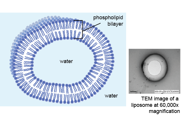
Cell surface (plasma) membrane.

Structure of membranes

  • The phospholipid bi-layer is visible using the electron microscope on a very high magnification (x100,000 -One hundred thousand)
  • Around 7 nm thick

Fluid mosaic model

  • Fluid: Because phospholipids and proteins can move around by diffusion - Phospholipids mainly move sideways within their own layer (monolayer) , while some proteins can move within the bi-layer. The fluidity of the bilayer is similar to that of olive oil
  • Mosaic: Pattern of scattered proteins when the membrane is viewed from above.
  • Model: Because we cannot magnify a cell enough to see the cell membrane, so we just have a model of what we predict the membrane looks like.

Features of fluid mosaic model

  • Double layer (bi-layer) of phospholipids
  • Individual phospholipid molecules move around their own mono-layers by diffusion.
  • Phospholipid tails point inward, forming non-polar hydrophobic interior
  • Phospholipid heads face outwards, into the aqueous (water-containing) medium that surrounds the membranes.
  • Some phospholipid tails are saturated and some are unsaturated (eg. cholesterol).
  • The more unsaturated they are, the more fluid the membrane is; this is because unsaturated fatty acid tails are bent, and therefore fit together more loosely.
  • Tail length also affects fluidity: the longer the tail, the more fluid the membrane is.
  • When temperature decreases, membranes become less fluid because there is less kinetic energy.
    Some organisms, such as bacteria and yeasts, who cannot regulate their own body temperature, respond by increasing the proportion of unsaturated fatty acids in their membranes to maintain fluidity.
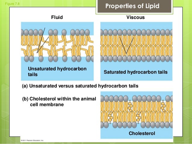
Unsaturated tails in the phospholipid bi-layer

Fluid mosaic model


Tuesday, November 24, 2015

Psychology: Development psychology: Transmission of aggression through imitation of aggressive models

Psychology: Development psychology: Transmission of aggression through imitation of aggressive models

Authors: Bandura et al. (1961)

Key term: Aggression

Background/context: Behaviorists believe that all behavior is learned, through classical conditioning (Pavlov - learned through association) and operant conditioning (Skinner - learned through reward and punishment). Watson (1923) classically conditioned 'Little Albert' to be scared of a white rat.
Behaviorists believed that we could only learn things that happened to us personally. But Bandura outlined observational learning - if behavior of a model (parent, teacher, etc) is observed then it will be copied (imitation learning). To test this theory Bandura designed an experiment trying to teach children aggression through observation.

Aim/Hypothesis: Children would reproduce aggressive behavior even if the model was no longer present
  1. If a behavior is observed it will be imitated.
  2. If a behavior is not observed it cannot be imitated.
  3. Boys will copy a male model more than a female model/ Girls will copy a female model more than a male model.
  4. Boys are more predisposed to show aggression than girls.
Method: Laboratory experiment - controlled observation 

Variables:
  • Independent variables:
  1. Three conditions:
    -Aggressive model group - 6 boys and 6 girls with the male model; 6 boys and 6 girls with the female model.
    -Non-aggressive model group - 6 boys and 6 girls with the male model; 6 boys and 6 girls with the female model.
    -Control group - 12 boys and 12 girls who saw no model at all.
  2. Sex of model
  3. Sex of child
  • Dependent variable: Number of behaviors out of 240 maximum in each response category.
bob doll study sampleIndependent variable conditions

Design: Matched pairs - Children were matched for pre-existing aggression levels of aggression, and independent groups as shown in 3 conditions above.

Participants and sampling technique: 
  • 72 children (36 boys and 36 girls) aged between 37(just over 3 years) to 69 months (5 years 9 months) from Stanford University nursery school.
  • Mean age was 52 months (4 years 4 months)
  • Probably opportunity sample
  • Quota sampling to achieve 12 participants in each sub-category
Apparatus: 
  • Room 1: Potato prints, picture stickers, table and chair, Tinker toy set, mallet and inflatable 5-foot bobo doll (adult-size)
  • Room 2: Fire engine, locomotive (train), doll, spinning top
  • Room 3: One-way mirror for observations, 3-foot bobo doll, mallet and peg board, two dart guns, tetherball with a face, tea set, 3 bears, cars, farm animals, crayons and coloring paper.
Bobo doll
Controls:
  • Children were matched for pre-existing aggression levels by the experimenter and nursery teacher independently rating 51 children on a scale of 0 - 5 (5 being very aggressive). There was very good agreement between the teacher and experimenter (0.89).
     -eg. A child rated as 5 (very aggressive) was matched with another child rated 5 (with one going to the 'aggressive' condition and one going to the 'non-aggressive' condition).
  • The toys in room 1, 2 and 3 were always the same and always in the same position when a child entered the room. 
  • The actions of the aggressive model was always the same, in the same order and for the same length of time
  • Observers watching through the two way mirror were unaware of which condition the child was in while observing to prevent bias.
  • The 20 minute session was divided into 5 second intervals, giving 240 response 'units'
  • Observers had a inter-rater reliability rate in the 0.9 range.
Procedure:
  1. Each child was shown to room 1. He/She played with the potato prints and stickers to settle in
  2. The child was taken to the other side of the room where they were shown to either of the two conditions for 10 minutes: aggressive or non aggressive.
     -Aggressive condition: Model would sit on, throw the bobo doll (Verbal aggressive: "throw him in the air") punch in the nose ("sock it on the nose"), hit the bobo doll with the mallet ("hit him down") and kick the bobo doll around the room ("kick him").
     -Non-aggressive condition: Adults assembled toy and did not interact with the bobo doll.
     -Control condition: No adult in the room.
  3. Child was then taken to room 2 for Mild aggression arousal (to annoy children and increase aggressive behavior). He/She played with the toys for around 2 minutes before the toys got taken away from the child and he/she was told they were not allowed to play with anymore as they were "the very best toys" and had been reserved for other children.
  4. Testing for delayed imitation. Room 3 (test room) contained non-aggressive and aggressive toys (eg. dart guns - aggressive gun play). The experimenter was in the room occupied with paperwork, while two observers watched through a two way mirror. Children were observed playing for the next 20 minutes.
Results:
  • Results were in 6 response categories for both female and male children:
    - Imitative physical aggression
    - Imitative verbal aggression
    - Mallet aggression
    - Punches bobo doll
    - Non-imitative aggression
    - Aggressive gun play
  • Imitative aggression: Physical and verbal aggression to that modeled in the procedure
  • Non-imitative aggression: New aggressive acts not demonstrated by model
  • Quantitative data
Results table

  • Significant results:
    Male imitative physical aggression with male model: 25.8
    Female imitative verbal aggression with female model: 13.7
    Male mallet aggression with male model: 28.8
    Male Non-imitative aggression with male model: 36.7
Findings:
  • Significantly more instances of aggression in the aggressive group than the non aggressive group.
  • Boys followed the male model more when it came to physical aggression.
  • Girls followed the female model more in verbal aggression.
     This may be because since a young age we are taught that men are meant to be more masculine.
  • Children in all groups, boys and girls, showed aggressive gun-play even though this was not observed in Room 1 (exposure) - non-imitative aggression.
Conclusion: Behavior that is observed is likely to be imitated - Bandura's 'observational learning' theory is supported. 

Strengths:
  • Laboratory experiment - High levels of control - Reliable, can be redone, EVs rare
     -Same toys in the same position for each child
     -Models did the same procedure for each condition for each child
     -All variables besides conditions in the IV controlled
     -Observers did not know which condition they were recording results for, so no bias.
  •  -Standardized procedure, can be repeated again to test for reliability of results
  • Valid - IV directly affects DV - Observation of models determining child's level of aggression
  • Quantitative data - Objective, can be analysed
     -Number of responses marked for each response category to show aggression in children clearly shows that the children who experienced the aggressive condition show more aggression.
Weaknesses:
  • Laboratory experiment - Lacks mundane realism - Not normal daily life activity
     -Children do not usually sit there and watch adults play without joining in
     -Children and model are strangers
     -Child and model do not interact
  • Quantitative data - We don't know why the children chose to be aggressive
  • Snapshot study - Results recorded immediately after experiment, which was also a single experiment - can one experience cause long term effects?
Ethics: 
  • Use of children in experiments is unethical because they cannot give consent
  • This experiment could have left long term psychological damage on the children
  • Children left the experiment psychologically different then before the experiment