Friday, August 30, 2019

Biology: Chapter 7: Transport in plants: Transpiration

Biology: Chapter 7: Transport in plants: Transpiration

Transpiration


  • Transpiration: Mesophyll cells in leaf have many air spaces between them filled with water vapor - Walls of the mesophyll cells are wet, and the water evaporates into these air spaces.
  • Air inside and outside of the leaf has direct contact through the stomata. If the water potential outside the cell is lower, then water diffuses out through the stomata.
  • Some water is used for photosynthesis but most is lost through evaporation this way.
  • When water evaporates out of the leaf more water is drawn to replace it, which comes from the xylem vessel.
  • The water moves down a concentration gradient from cell to cell in two possible pathways:
  • Apoplastic: Without entering the cells, water travels between the cell walls.
  • Symplastic: Water travels cell to cell via plasmodesmata.
Xylem tissue


  • Made from cells joined end to end.
  • Vessel elements and tracheids: Involved in transport of water.
  • Sclerenchyma fibers: Elongated cells with lignified walls to support plant. 
  • Dead at maturity.
Xylem vessels:

  • Vessel elements begins as a normal plant cell with lignin in its wall. 
  • As lignin builds around cell, cell and its contents die leaving a empty space inside the cell.
  • Lumen: Empty space in the cell.
  • Plasmodesmata of these original cells form gaps with no lignin in the cell walls. These form gaps in the xylem vessel wall.
  • Pits: Gaps in the wall and throughout the xylem vessel. Made of freely permeable cellulose cell wall. Link with pits of neighboring cells so water can pass freely from one cell to the next.
  • End walls break down and form a continuous tube.
  • This continuous non-living tube is the xylem vessel and can be up to several meters long.
Transpiration

  • When water leaves the xylem tube at the leaves it creates a lower hydrostatic pressure at the top of the tube than the bottom.
  • The pressure causes the water to move up the tube in continuous columns.
  • Strong lignified walls stop vessels from collapsing inwards due to pressure.
  • Mass flow: Movement of water up the xylem vessels as a body of liquid, as a result of cohesion and adhesion.
  • Cohesion: Attraction between water molecules.
  • Adhesion: Attraction of water to cellulose and lignin in the xylem vessel.
  • Small diameter and pits of the vessel help prevent air bubbles from forming, which would change the pressure and disrupt mass flow. 
  • Pits allow water to move out into neighboring living cells and prevent air locks.

Friday, January 29, 2016

Psychology: Physiological psychology: Olfactory (smell) module facial attractiveness

Psychology: Physiological psychology: Olfactory (smell) module facial attractiveness

Authors: Dematte et al. (2007)

Key terms: Smells and facial attractiveness

Background/ Context:
  • Facial attraction is a socially important cue that has been researched in depth.
  • Studies show smell is very important for animals in determining their mate.
  • Therefore, is attractiveness determined by other cues such as olfaction?
  • Olfaction: Sense of smell.
  • This study focuses on female participants because it is said that females are more sensitive to olfactory cues than men, and might rely more on olfactory cues when it comes to mating behaviour.
Aim/ Hypothesis:
  • Discover whether olfactory cues affect people's judgements of attractiveness.
  • Specifically: Whether pleasant odors increase male attractiveness, and whether unpleasant odors decrease male attractiveness.
Method: Laboratory experiment (including minor questionnaire)

Variables:
Independent variables:
  1. Three conditions: Pleasant odors (Geranium and Gravity), Unpleasant odors (Synthetic body odor rubber) and a neutral odor (clean air).
  2. High facial attractiveness, low facial attractiveness.
  • Dependent variable: Ratings of attractiveness from 1 (least attractive) to 9 (most attractive).
Design: Repeated measures and counter-balancing.
  • Counter-balancing so each 40 face was presented with a different odor condition.
  • 3 blocks of experiment sessions with a 5 minute break between each. 2nd and 3rd session replicated the 1st.
  • Each block presented all 40 faces; divided into 4 sets of 10 faces. 
  • Each set of 10 faces were divided into 5 attractive and 5 unattractive.
  • No two odors were presented together.
  1. Set 1: 10 faces with clean air, geranium and body odor.
  2. Set 2: 10 faces with clean air, gravity and rubber.
  3. Set 3: 10 faces with clean air, geranium and rubber.
  4. Set 4: 10 faces with clean air, gravity and body odor.
  • Order of presentation randomized.
  • Whole experiment lasted 50 minutes per participant.
  • 120 trials in total for block 1. Same procedure repeated for a 2nd and 3rd time.
  • Trials were randomized in each block. 
Participants and sampling technique:
  • 16 female students from Oxford University, UK.
  • Aged 20-34.
  • None knew the purpose of study.
  • Completed confidential questionnaire to ensure they all had a normal sense of smell - no olfactory disorders, asthma, no flu. normal or corrected (eg. glasses) vision. This was to make sure no other variables other than odor affected their judgement.
Apparatus:
  • Questionnaire to determine health (eg. olfactory or respiratory) problems.
  • 40 male faces (13 x 17 cm) from a standardized face data base (previously categorized into attractive and unattractive faces).
  • 4 odors: 2 pleasant (geranium and gravity), 2 unpleasant (synthesized body odor and rubber) and control with no odor (clean air). 
  • Olfactometer: Computer controlled machine to deliver odors. 
  • Computer to view the male face images.
Controls;
  • Questionnaire before study to make sure participants can detect smell normally ("Are you currently suffering from a cold/flu?", "Do you suffer from asthma or any air-born allergy?") - to ensure validity and ability to generalize.
  • Presentations of faces and odors were counterbalanced - each face was presented with each odor to remove order effects and increase validity.
  • Presentations of pleasant and unpleasant odors were counterbalanced to also prevent order effects.
  • Presentation time was controlled at 500 milliseconds (Half a second) to increase validity.
  • Time for tone presentation and release of odors was standardized.
  • Odor intensity standardised for each participant because each odor has a different intensity; Odors were diluted to ensure not one smell was more intense than the others: Body odor 0.33%, Geranium 1%, Gravity 0.5% and Rubber 1.2%, which was confirmed during the pilot study.
Procedure:
  1. Participant sat on a chair 70 cm from the computer screen. A chin rest was used to keep their head stable.
  2. Participants were told to look at the cross on the screen.
  3. Participants were told to exhale when they heard a quiet tone.
  4. Participants were told to inhale through their nostrils when they heard the loud tone.
  5. Half a second after participants inhaled the 4 odors or clean air were delivered through the olfactory machine.
  6. A second after the odor had been delivered a face would appear for 500 milliseconds (half a second) - So participants would not have time to analyse and examine the face. When the face disappeared clean air was delivered, cleansing the room of any odor.

Thursday, January 28, 2016

Biology: Chapter 7: Transport in plants: Parts of plants

Biology: Chapter 7: Transport in plants: Parts of plants


Flowering plants (Angiosperms)
  • Cotyledon: First part of seedling to emerge from soil when it germinates.
  • Monocotyledons (Monocot): Plants that have only one cotyledon. Usually have long narrow leaves and flower parts in multiples of 3 eg. grass.
  • Dicotyledons (Dicot): Plants with 2 cotyledons. Usually have leaves with blades and stalks eg. sunflowers. All plants in this chapter are dicotyledons.

Parts of the plant

Cortex: 
  • Outermost layer of root and stem tissue in plants. 
  • Between Epidermis and Endodermis.
  • Composed mostly of parenchyma and specialized cells.
Pith: Central region of stems.

Epidermis: 
  • Outermost layer of whole plant structure.
  • Covers the whole plant.
  • One cell thick. Closely packed, without chloroplasts or space between cells.
Endodermis:
  • Innermost layer of cortex.
  • Surrounds vascular bundle.
  • One cell thick.
  • Controls entry of water and substances into vascular bundle by Casparian strip.
Mesophyll:
  • Leaf cells
  • Specialised parenchyma cells found between lower and upper epidermis of the leaf.
  • Specialised for photosynthesis: contain chloroplasts. 
  • Palisade mesophyll: Column shaped. Near the upper surface of leaf to recieve more sunlight. Contain more chloroplasts than spongy mesophyll.
  • Spongy mesophyll: Irregular sponge shapes. Underneath palisade cells. Have many large air spaces between the cells to let gas from the stomata diffuse around cells.
Parenchyma:
  • Thin walled cells.
  • Living at maturity,
  • Used as packing tissue.
  • Metabolically active - many functions.
  • Can specialize. 
  • For example: Storage of starch. When turgid, help support plant and prevent wilting. Air spaces between these cells allow gas exchange. Water and mineral salts transported through walls and living contents of cell. 
  • Contain chloroplasts in leaves - specializes to become palisade and spongy mesophyll tissue.
  • Form cortex in roots and stems.
  • Form pith in stems.
Collenchyma:
  • Modified form of parenchyma with extra cellulose deposited at ends of cells, providing extra strength.
  • Living at maturity.
  • Midrib of leaves contains collenchyma.
Sclerenchyma:
  • Thick secondary cell walls.
  • Contain lignin for extra strength.
  • Lignin: With cellulose, main component of wood. Water proof and strong.
  • Dead at maturity.
  • Cannot increase in length.
  • Found anywhere in plant.
  • Provide support in plants.
Pericycle:
  • One to several cells thick.
  • Between the endodermis and vascular bundle.
  • In roots, is one cell thick, and new roots can grow from this layer by meristematic cells.
  • Meristematic cells: Plant stem cells.
  • In leaves, formed by sclerenchyma cells, which is dead and lignified for extra strength.
Stele: Contains vascular bundle in roots and stems.

Vascular tissue:
  • Contain the types of xylem and phloem systems.
  • Tubes for transportation of fluids.
  • Xylem and phloem found in vascular bundles.
  • Outsides made of sclerenchyma fibres which provide exta support.
Xylem:
  • Long distance transport of water and mineral salts. 
  • Xylem vessel elements: Tubes made of dead cells. Reinforced with lignin. 
  • Dead at functional maturity.
Phloem:
  • Long distance transport of organic compounds eg. Sucrose.
  • Living cells.
  • Have a companion cell alongside the tube.
  • Contain tubes called sieve tubes 
  • Sieve tube elements: Living cells that make up the sieve tubes.


Monday, January 25, 2016

Psychology: Physiological psychology: Taxi drivers brain activity

Psychology: Physiological psychology: Taxi drivers brain activity

Authors: Maguire et al. (1997)

Key term: Taxi drivers

Background/ Context: The invention of technology such as PET and MRI scanners has led to many studies on brain regions and activity. Topographical memory is the knowledge of landscapes and the spatial relationship between them - a cognitive (mental) map we have inside our heads. Maguire's study is on semantic topographical memory and what brain regions are responsible for it.
PET: Positron Emission Tomography.
Semantic memory: General world knowledge we have

Aim/ Hypothesis;

  1. Confirm findings of other studies that specific brain regions are responsible for semantic topographical memory retrieval
  2. Determine which specific region of the brain is responsible for landmark knowledge when location information is not needed (topographical memory without sequencing).
  3. Study topographical and non-topographical semantic memory to see if there is brain region that is activated for both
Method: Laboratory experiment with pre-study questionnaire
  • Participants filled out a questionnaire asking them about:
    -Areas of London they were familiar with.
    -Films they were familiar with from a list of 150 films from 1939 to the present day.
    -20 world famous landmarks participants could visualize but had never visited.
  • This would let experimenters choose the most appropriate routes, landmarks and films they wanted the participants to describe.
  • Pilot study done with non-participating taxi drivers before this experiment to make sure the tasks and equipment were fit for the study.
Variables:
Independent variables
  • Main variables: Topographical and Sequencing.
  • The participants completed 6 tasks, each done twice. However only 5 of those tasks are used in this study.
  1. Baseline task - Repeat two sets of 4 digit numbers (to compare simple level of activity to the 4 other tasks).
  2. Describe the shortest route from one point to another in the City of London (same route for all participants, one they were familiar with) (topographical and sequencing).
  3. Describe a landmark in terms of features, appearance, etc (Same landmark for all participants, one they all knew) (topographical and non-sequencing).
  4. Describe the plot of a famous film between two given points of the film. (familiar to all participants) (non-topographical and sequencing).
  5. Describe individual frames of some famous films eg. imagery, characters, etc. (familiar to all participants) (non-topographical and non-sequencing). 
Dependent variable: Region/s of brain activated.

Design: Repeated measures - Factorial design
  • Orders of tasks randomized for each participant and counterbalanced against each other.
Participants and sampling technique:
  • 11 right handed male taxi drivers.
  • Minimum of 3 years experience driving, average was 12.5 years.
  • Average age 45 years.
Apparatus:
  • London route from Grosvenor Square to the elephant and castle.
  • World famous landmarks
  • Films with still frames
  • Siemens PET scanner
  • H2 15 O bolus (radioactive isotope for scanning) and saline flush to remove isotope from body.
Controls:
  • Landmarks chosen were places participants never visited in person, so they were not sequential memories.
  • Films chosen had been watched by participants at least 5 times, so they were all familiar with them.
  • Scanner procedure and injections given were the same for each participant.
  • Pilot study done to ensure all tasks are fit for purpose.
Procedure:

  1. Participant filled out pre-study questionnaire.
  2. Participant arrived at the laboratory and cannula (tube for delivering fluid) was inserted in his arm. Participant was blindfolded and placed in scanner.
  3. Participant received H2 15O bolus injection through cannula (which takes 20 seconds to enter bloodstream). This was followed by a saline flush (which also takes 20 seconds). 
  4. Scan frame time was 90 seconds (H2 15O has a half life of 2 minutes). 1 of the 5 tasks was presented and the participant had to give a description of the route, landmark, film plot or frame, which was digitally recorded. MRI was also taken for each taxi driver,
  5. After the task was completed their would be an 8 minute wait before another task was done.
  6. Steps 3, 4 and 5 repeated 12 times (2 times for each study including another task for another study not in this paper). 
  7. Participant was debriefed and study was complete.
Data: Quantitative data - rCBF (Regional cerebral blood flow) would flow to areas of brain activated, which would be recorded by the PET scanner, interpreted and analysed. 3D images of the PET scanner show exactly which brain region is involved in each process.

Findings:
  • No difference in amount of speech per participant (around 50 seconds).
  • Very few differences in route chosen by participants.
  1. Routes (T+ S+): Right hippocampus, parahippocampal gyrus, cerebellum, media parietal lobe, posterior cingulate cortex, extrastriate regions.
  2. Landmarks (T+ S-): Parahippocampal gyrus, posterior cingulated cortex, medial parietal lobe, occipito-temporal region, cerebellum.
  3. Films (plot and frames - T- S+ and T- S-): Left frontal region, cerebellum, middle temporal gyrus, left angular gyrus.
  • Cerebellum activated in all activities
  • The parahippocampal gyrus and medial parietal lobe were used for both topographical activities. However, the right hippocampus was only activated in the route activity - topographical and sequencing.
Conclusion:
  • Regions of topographical semantic memory similar as those in previous studies.
  • Right hippocampus used for sequential route planning.
Strengths:
  • Laboratory experiment - High level of control - pilot questionnaire, routes, landmarks and films chosen - easily replicated to test for reliability.
  • Laboratory experiment - High level of control - confidence the type of tasks directly affected brain function - IV directly affects DV - baseline task, type of tasks.
  • Technological equipment - lack of human error 
  • Testing physiological aspects (brain function) - can be generalized to wider population, as majority of humans have similar brain structures.
Weaknesses:
  • Laboratory experiment - Artificial setting - Participants were tested whilst undergoing a PET scan, which is not a normal setting for recalling memory, therefore this experiment may not have ecological validity
  • Laboratory experiment - Lacks mundane realism - Participants were blindfolded and asked to recall memories during a PET scan, which is not a normal task people do in daily life.
  • Can't be generalized - Participants were all taxi drivers - It is possible the right hippocampus in sequential topographical memories is only crucial in taxi drivers.

Wednesday, January 20, 2016

Biology: Chapter 6: Nucleic acids and protein synthesis: Protein synthesis

Biology: Chapter 6: Nucleic acids and protein synthesis: Protein synthesis

  • Protein synthesis: Process in which the bases on the DNA molecule is used to code for the sequence of amino acids in a polypeptide.
  • Sequence of amino acids determine the 3-D folding of the protein and therefore it's function.

Transcription

  • A section of the DNA (gene) has the instructions for creating a mRNA (messenger RNA) molecule.
  • Promotor: Signals the start of a gene.
  • Terminator/ Stop sequence: Signals the end of the gene.
  • RNA polymerase: Binds to the promotor and when the DNA uwinds initiates RNA synthesis.
  • Sense strand: DNA strand with the same code as the mRNA.
  • RNA polymerase starts RNA synthesis as the DNA unwinds at the promotor. 
  • Moves down the DNA strand in a 5' to 3' direction, until it reaches the base code for the stop sequence at the end of the gene, where the newly formed RNA transcript peels off the template strand.
  • DNA rewinds back into the double helix after the RNA synthesis.
  • The mRNA breaks away from the DNA and leaves the nucleus through the nuclear pores into the cytoplasm.
  • mRNA (messenger RNA): RNA transcript created in translation.
  • Template strand: DNA strand that the mRNA uses as a template for RNA synthesis - the antisense strand.
Simple diagram of transcription.

Translation

  • Process of building chain of amino acids following code on mRNA molecule made in transcription.
  • mRNA leaves nucleus through nuclear pores into the cytoplasm.
  • Ribosome (made of rRNA - ribosomal RNA) clamps around the mRNA.
  • Ribosomes: Responsible for protein synthesis.
    Have 2 units - large subunit and small subunit, which clamp around the mRNA.
    The large subunit has 3 sites - A siteP site and E (exit) site.
    These are for tRNA (transfer RNA) to come in and bond to it's complementary codon.
    Two tRNA molecules can be in a ribosome at one time.
    Ribosome.
  • Codon: Groups of 3 bases which code for an amino acid
  • Anti-codon: 3 bases on a tRNA. Forms a bond with it's complementary codon on the mRNA strand in the ribosome.
  • tRNA (transfer RNA): Made from a single strand of RNA, twisted to form a 3 leaf clover shape. Has a anti-codon (base triplet) on one end, and amino acid attachment site on the other.
    tRNA molecule.
  • The tRNA bonds to an amino acid specific to it's anticodon and carry it to the ribosome, where it forms hydrogen bonds with it's complementary codon on the mRNA strand.
  • 2 tRNA molecules are in the ribosome at one time. The ribosome cuts the bonds of the amino acid and the tRNA in the P site, and the amino acid forms a peptide bond with the amino acid on the tRNA in the A site, forming a amino acid chain.
  • The ribosome then moves down the mRNA chain, in a 5' to 3' direction. 
  • The tRNA that was previously in the P site moves to the E site, and is released from the ribosome. 
  • The tRNA in A is now in P, and a new tRNA enters the A site
  • The ribosome again cuts the bond between the amino acids and tRNA in the P site, which again form a peptide bond with the amino acid on the tRNA in the A site
  • The process continues, forming a polypeptide chain.
    Ribosome in translation.
  • start codon signals the start of translation in the ribosome - usually AUG
  • When the stop codon is reached on the mRNA strand - usually UAG, UAA or UGA, the ribosome accepts a release factor, which allows the ribosome to break the bond between the last tRNA and it's now polypeptide chainreleasing the polypeptide chain, which will fold into secondary, tertiary and quaternary structures according to it's amino acid sequence.
  • The ribosome and tRNA seperate from the mRNA chain.
  • There are 20 different types of amino acids present in the cytoplasm.
  • Many ribosomes work on the mRNA strand at one time, so multipe copies of the polypeptide can be made efficiently.
    Translation diagram.
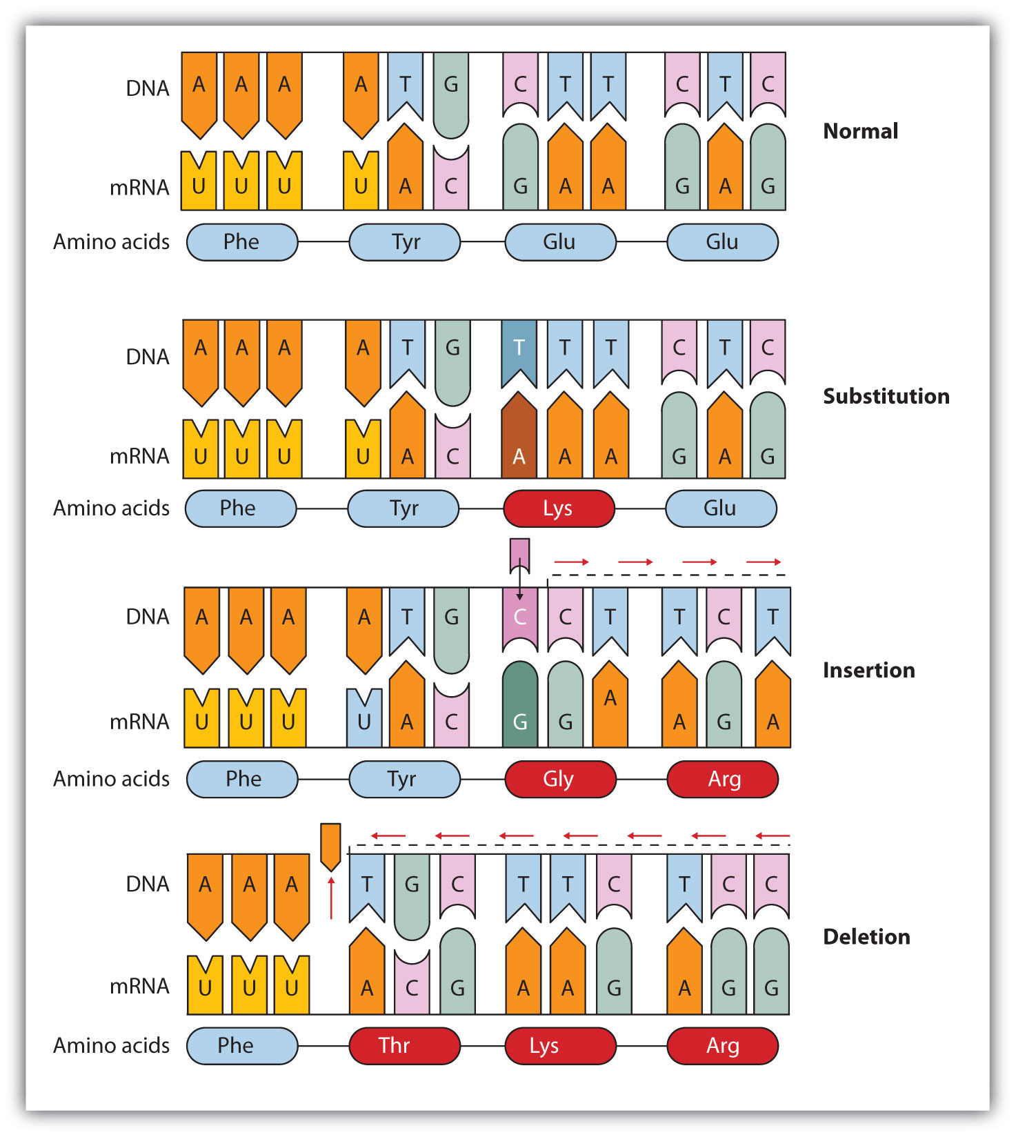
Mutations

  • Changes in the genetic material.
  • Change of bases in the code, which can lead to a new gene (alleles).
  • Alleles: Different types of the same gene (Blue or brown eyes).
Point mutations - Changes in just one base pair
  • Substitution: Replacement of one nucleotide and it's complementary base in the DNA strand. Leads to alteration in amino acid sequence, which can hinder protein function eg. Sickle cell anemia - glutamic acid replaced with valine.
  • Insertion/Deletion: nucleotide pair inserted or removed in the DNA strand. Can result in an extra or missing amino acid. May alter reading frame (codon groups) - Frameshift mutation, which creates a protein that is almost certainly unable to function.
DNA mutations.

Monday, January 18, 2016

Biology: Chapter 6: DNA and Nucleic acids: DNA replication

Biology: Chapter 6: DNA and Nucleic acids: DNA replication

  • In 1953, James and Watson Crick used the results of work by Rosalin Franklin to develop a model of the structure of DNA.
  • Semi-conservative replication: Method of copying where half of the original molecules are kept (conserved) in the new molecules.
    Simple diagram of semi-conservative replication in DNA.
  • Enzyme Helicase unzips parental double helix, separating the two strands.
  • Replication fork: Where the DNA strands are separating.
    Replication fork.
  • Single-strand binding proteins: Keep the two parental strands separated by temporarily hydrogen bonding to the bases.
  • Primase: Adds RNA primer to DNA strand
  • RNA primer: Initiate DNA synthesis by DNA polymerase
  • DNA polymerase: Builds a new strand by assembling the nucleotides to the parental strand. Nucleotide bases form the specific bonds with its complementary base on the parental strand, therefore it is almost impossible to make a mistake. Only works in a 5' to 3' direction.
  • Since DNA polymerase can only work from a 5' to 3' direction, one strand can be built continuously as the replication fork progresses. 
  • Leading strand: Strand that is built continuously from from 5' to 3'.
  • However, one strand will have the replication fork progressing towards 5'. This strand will grow in the overall direction of 5' to 3' by the addition of short fragments as the replication fork progresses. 
  • Lagging strand: 3' to 5' strand that needs to be built in fragments.
  • Okazaki fragments: Fragments used to build the 3' to 5' strand.
  • Another DNA polymerase replaces RNA primer with DNA.
  • DNA ligase: Joins okazaki fragments together to growing strand.
DNA replication diagram.

Proofreading DNA
  • DNA polymerase proofreads each nucleotide and removes incorrectly paired nucleotides.
Repairing DNA
  • DNA molecules that are exposed to harmful chemical and physical agents eg. Tobacco, UV rays can change nucleotides, which can affect encoded genetic information. However, the DNA has some ways of fixing it.
Nucleotide excision repair:
  • Nuclease enzyme: Cuts damaged DNA strand at it's 2 points.
  • Repair synthesis by DNA polymerase fills gap
  • DNA ligase: Seals the phosphate sugar backbone together.

Friday, January 15, 2016

Biology: Chapter 6: Nucleic acids and protein synthesis: DNA and RNA structure

Biology: Chapter 6: Nucleic acids and protein synthesis: DNA and RNA structure

Nucleotides aka Nucleic acids

  • Smaller molecules that DNA and RNA are made up of.
  • Polynucleotides: Long chains of nucleotides eg. DNA and RNA.
  • Composed of 3 components:
    -Pentose sugar
    -Nitrogen-containing base
    -Phosphate group
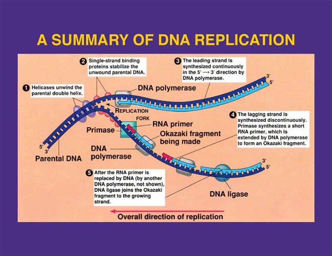
Structure of nucleotide.
  • Pentose sugar: Sugar with 5 carbons. Can be either Ribose (in RNA) or Deoxyribose (in DNA.
  • Deoxyribose: Missing an oxygen on it's second carbon primer.
Nitrogen-containing bases aka Nitrogenous bases
  • Purine bases: Double ring structure - Adenine, Guanine.
  • Pyrimidine bases: Single ring structure - Thymine, Cytosine, Uracil.
  • DNA contains the 4 bases A-T and C-G, but never Uracil.
  • RNA contains the 4 bases A-U and C-G, but no Thymine.
Structural formulae of the nitrogenous bases.

ATP: Similar structure to a nucleotide. Adenosine (Ribose sugar with the base Adenine) can be combined  to 1,2 or 3 phosphates to make Adenosine Monophosphate (AMP), Adenosine Diphosphate (ADP) or Adenosine Triphosphate (ATP).

Polynucleotides
  • Polynucleotide: Many nucleotides linked together in a chain
  • Formed during interphase of the cell cycle.
  • Sugar-phosphate backbone: Alternating sugars and phosphates of the nucleotides linked together with the bases projecting sideways.
  • Phosphodiester bonds: Covalent sugar-phosphate bonds.
  • Phosphodiester bonds link the Carbon 5 of one sugar to the Carbon 3 of the next.
  • The polynucleotide is said to have 3' and 5' ends: the end where the Carbon 3 has nothing to link is called the 3' end, while the end where the Carbon 5 has nothing to link to is called the 5' end.
  • Bases can be purines or pyrimidines: Purines only link with pyrimidines, but it is even more precise.
  • In a DNA molecule, there is only enough room between the two sugar-phosphate backbones for 1 purine and 1 pyrimidine molecule, so a pyrimidine can only bond with a purine, since purines are larger.
  • Adenine and Thymine form double hydrogen bonds, whereas Guanine and Cytosine form a triple hydrogen bond, therefore only pairs possible are A-T and C-G.
  • Complementary base pairing: The precise pairing of bases due to hydrogen bond and structure size (purine/pyrimidine) : Adenine and Thymine (A-T), and Cytosine and Guanine (C-G).
Animation of DNA replication and the DNA strand.

  • Double helix: Two strands wrapping around each other - the 3D shape DNA molecules form.
  • Hydrogen bonds linking the bases easily broken, which happens during DNA replication and protein synthesis. 
  • RNA molecules are only single stranded.