Sunday, December 27, 2015

Psychology: Developmental Psychology: Case study 3: Infant facial preference

Psychology: Developmental Psychology: Case study 3: Infant facial preference

Authors: Langlois et al. (1991)

Background/Context: Is beauty in the eye of the beholder? Do we judge books (and people) by their cover?
Langlois believes that there is a universal standard of beauty that people judged facial attractiveness with, and both adults and infants judge attractive adults and infants more favorably, treating them better than those who are unattractive. 
Young infants have not yet been influenced by media or culture, so researching on infants would prove whether there is a universal standard of attractiveness.

Aim/hypothesis: 
  • Study 1:
    -Replicate previous results (preference for attractive) with adult female faces.
    -See if adult male faces produce the same results (preference for attractive).
    -Investigate where order of presentation of male/female affects preferences.
  • Study 2: See if same results are achieved with non-white faces.
  • Study 3: See if same results are achieved with faces of babies.
Method: Laboratory experiment

Variables:
  • Independent variables:Study 1-Attractive and unattractive white female faces-Attractive and unattractive white male faces-Infants who saw either of two conditions: All men then all women (or all women then all men) and alternating men and women.

    Study 2: Attractive and unattractive black female faces.

    Study 3: Attractive and unattractive male and female infant faces.
  • Dependent variable: Fixation time (in seconds) - length of time an infant looks at a face. Each face was presented twice (once on the left, and once on the right of the screen) so the mean time for each face was calculated and agreement between the two observers could be checked.
  • Attractiveness of faces judged by people on the 5-point Linkert scales.

Thursday, December 17, 2015

Psychology: Developmental psychology: Analysis of a phobia of a 5 year old boy

Psychology: Developmental psychology: Analysis of a phobia of a 5 year old boy

Author: Freud (1909)

Key term: Little Hans

Approach: Development psychology; psychodynamic perspective

Method: Case study and longitudinal method

Freud's psychodynamic approach:
  • Our behaviours and feelings when we grow up (including psychological problems) are caused by childhood experiences.
  • All behaviour is determined.
Personalities are made up of 3 parts: The ID (instinctive drive), ego and superego
  • ID: Biological aspects of the personality. Consists of two forces:
          Eros: Life force - Instinct that drives us to do biological things - sex drive, eating, etc. Mainly revolves around libido (sex drive).
          Thanatos: Death force- Instinct of self-destruction.
    ID is chaotic and totally unreasonable (we cannot randomly have sex with people when we feel like it).
  • Ego: Mediates between unrealistic ID and external real world. Decision making component of personality. Works out realistic ways of satisfying ID.
    "Part of the ID which has been modified by the direct influence of the external world" - Freud.
    Similar to ID, seeks pleasure and avoids pain but using a realistic strategy.
  • Superego: Values and morals of society by influence of parents or role models. Strive for perfection. Develops at around the age of 3-5 during phallic stage of psychosexual development. Two systems:
        Conscious: Punishes the ego through feelings of guilt.
        Ideal self: Imaginary picture of how you ought to be; career aspirations, how to treat other people and how to behave as a member of society.
  •  The ego and superego are largely determined by parental values and how you were brought up.
Freud's psychosexual development theory:
  • Freud believed life was built around tension and pressure, which was due to the libido.
  • What develops is the way sexual energy accumulates and builds up and is discharged as we mature biologically. 
  • Each psychosexual stage has a particular conflict that must be resolved before continuing to the next stage. Some people may not be able to leave one stage and continue on to the next. This can be because the needs of the stage have not been met, causing frustration, or the pleasure is so good they don't want to leave, called overindulgence. 
  1. Oral - The mouth - sucking, swallowing, etc. Ego develops.
  2. Anal - The anus - withholding or excreting feces. 
  3. Phallic - The penis or clitoris - masturbation. Superego develops.
  4. Latent - Little or no sexual motivation.
  5. Genital - The penis or vagina - Sexual intercourse.
Freud's Oedipus complex
  • Oedipus - Greek myth where Oedipus, a young man, kills his father and marries his mother.
  • The Oedipus complex is when a 3 - 5 year old boy develops sexual feelings for his mother, and therefore wants to get rid of his father so he can have his mother all to himself.
  • This is usually resolved by the boy imitating his father's masculine type behaviours, called identification.
  • Happens during phallic stage.
  • Fred states that every boy goes through this complex to a certain extent.

Little Han's phobia case study

  • 5 year old Jewish boy from Vienna, Austria.
  • Phobia of horses
  • Primary aim was to treat the phobia.
  • Freud did not work directly with Hans. his father gave Freud all the details of Hans behavior when he suspected Hans was suffering from the Oedipus complex.
  • At 3, Hans was very obsessed with his 'widdler' (penis), and also those of other people. He once asked his mother "Mommy, do you also have a widdler?". He played with it regularly until his mother threatened to call the doctor to get it cut off, which made him get castration anxiety. 
  • At the same time, Hans saw a horse collapse and die in the street, and was very upset.
  • At 4, Hans developed a fear of horses, specifically a fear that a white horse would bite him. 
  • At the same time a conflict developed between him and his father. Hans had developed a habit of getting in to his parents bed in the morning to cuddle his mother. However, his father began to object and banned him from continuing.  
  • Hans phobia worsened to the point that he would not leave the family house and he also suffered attacks of generalised anxiety.
  • The anxiety Hans had was actually castration anxiety triggered by his mothers threat to cut off his "widdler" and fear of his father caused by his banishment from the parental bed. 
  • Hans reported having the following dream; "In the night there was a big giraffe in the room and a crumpled one: and the big one called out because I took the crumpled one away from it. Then it stopped calling out: and I sat on top of the crumpled one."
  • The giraffes in Hans dream represent his parents. The large giraffe is his father, and him crying out represented him objecting Hans. It's erect neck could have also been a penis symbol. The crumpled giraffe was his mother, and the crumpling represented her genitals. 
  • When Hans was 5 his phobia of horses lessened, initially becoming limited to only white horses with black nose bands, then disappearing altogether.
  • The horses represented Hans father, especially white horses with black nose bands, because they looked like his moustached father. Horses also have large penises, which make a good father symbol.s
  • The end of the phobia was marked by two fantasies:
  • Hans fantasised that he had several children. When his father asked who their mother was Hans replied "Why Mummy, and you're the granddaddy"
  • This children fantasy represents a friendly resolution of the Oedipus complex, where he replaces his father but still keeps him in the family with the role as grandfather.
  • The next day, Hans fantasised that a plumber had come and removed his bottom and penis, replacing them with new and larger ones
  • The plumber fantasy represents identification with the father. Hans can see himself growing a large penis like his fathers and becoming like him.
Conclusion
Hans had a phobia of horses because he was suffering from castration anxiety and was going through the Oedipus complex. Hans dreams and fantasies helped express this conflict and eventually he resolved his phobia and Oedipus complex by identification by fantasising of himself taking on his fathers role and placing his father in the role of grandfather.

Strengths

  • Case study - Lots of data can be collected because it is the study of one boy, so it is more valid.
  • Natural observation - Hans was in his own house and not in any artificial setting, so there is a high level of ecological validity and mundane realism.
Weaknesses
  • Unscientific - Freud's theory is considered unscientific because there is no evidence to back his theory.
  • Generalization - Since it is the study of only one boy, we cannot generalize the oedipus theory to every boy.
  • Bias - Freud published his theory before doing this case study, therefore he could have been bias. Hans father was a fan of Freud and believed in his theory, therefore he could've only told details that correlated to the study.
  • Invalid - There could be many other reasons Hans had a phobia of horses. For example, when he was 3 he saw a horse fall in the street and die.


Tuesday, December 15, 2015

Biology: Chapter 5: Mitotic Cell cycle: Mitosis

Biology: Chapter 5: Mitotic Cell cycle: Mitosis


  • Part of a precisely controlled process called the cell cycle.
  • Nuclear division: Splits the cell's nucleus into two daughter nuclei that are genetically identical and contain the same number of chromosomes as the parent cell.

Cell cycle

  • Regular sequence of events between one cell division to the next.
  • Three phases: Interphase, nuclear division (mitosis) and cell division (cytokinesis)
Interphase:
  1. G1 phase (Gap 1): Cell grows to normal size after cell division. Carries out normal function eg. Protein synthesis. At the end of G1, the cell receives a signal telling it whether or not it will divide. If a cell receives the go signal to divide, it will continue into the S phase.
  2. S phase (synthesis): DNA in the nucleus replicates - two sets of each chromatid that can form chromosomes.
  3. G2 phase (Gap 2): Cell continues to grow and the new DNA is checked for any errors and are repaired. Preparations made for division eg. Increased production of Tubulin, which is used to make spindle.
Nuclear division (mitosis):
  • Nucleus divides into two genetically identical daughter nuclei
  • Growth temporarily stops.
Cell division (cytokinesis):
  • After the two new nuclei move to the opposite poles of the cell, the cell splits into two by constricting the cytoplasm between the two nuclei
  • Cleavage furrow: Constriction of the cytoplasm between two nuclei.
  • Creates two new identical cells.
  • In plant cells, vesicles carry building materials (eg. cellulose) from the golgi body and fuse together to form a cell plate between the two nuclei.
Cell cycle diagram

Mitosis

  • Four phases: Prophase, Metaphase, Anaphase and Telophase (PMAT)
Prophase:
  • Chromatin coil up and chromosomes start to appear
  • Nuclear envelope, nucleolus and organelles 'disappear' - break up into smaller vesicles
  • Centrosomes replicate and start moving to opposite poles where they form the poles of the spindle fibre.
Metaphase:
  • Centrosomes at opposite poles help organize production of spindle microtubules.
  • Chromosomes line up across the middle of the spindle, attached by their centromeres.
Anaphase:
  • Spindle fibre attached to the centromeres of the chromosomes pull the two chromatids to separate poles, centromere first.
Telophase:
  • Chromatids reach the pole of their spindle and uncoil (each one contains 1 DNA molecule, which replicates itself during interphase for the next cell division)
  • Nuclear envelope and nucleolus reform around the chromatin
  • Remaining spindle fibre breaks down
  • Cytokinesis starts 

Significance of mitosis
  • Growth: Two daughter cells are identical and have the same number of chromosomes - allow for multicellular growth from a unicellular zygote. Can occur over the entire body or in specific regions.
  • Repair and replacement of cells: Dead or faulty cells can be replaced by identical cells.
  • Asexual reproduction: Production of new genetically identical individuals by a single parent organism. Can occur in unicellular organisms eg. Amoeba or in some plants (budding)
  • Immune response


Friday, December 11, 2015

Biology: Chapter 5: Mitotic Cell cycle: Components of a cell cycle

Biology: Chapter 5: Mitotic Cell cycle: Components of a cell cycle

  • Cells reproduce by dividing and passing on copies of their genes to 'daughter' cells. 
  • This is done through nuclear and cell division.

Chromosomes

  • Condensed form of chromatin that occurs only during mitosis. Chromatin during normal cell activity (interphase). Consist of 2 identical sister chromatids connected by a centromere. 
  • Each chromatid contains 1 DNA molecule - chromatids in chromosomes are identical to one another.
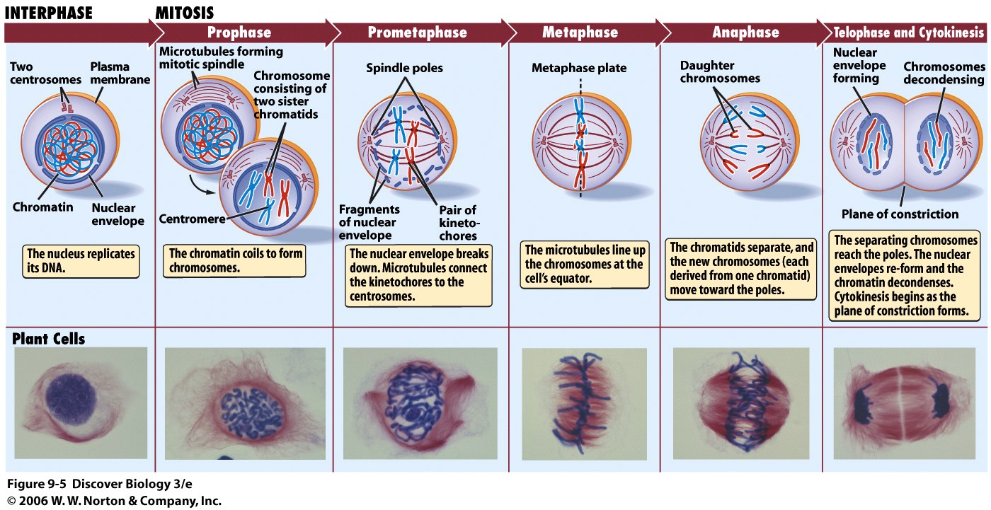
Chromosome during Anaphase
  • DNA: Molecule of inheritance. Made up of a series of genes. Only 2nm wide, but total length of the DNA in all the 46 chromosomes in an adult is approximately 1.8 meters. DNA is wound around histones precisely to keep them from being tangled. 
  • Gene: 1 unit of inheritance. Codes for 1 polypeptide that is involved in a specific aspect of the functioning of the organism.
  • Histone: Protein that DNA wounds around. Proteins are basic (alkali) which means they can easily interact with acidic DNA.
  • Nucleosome: DNA wrapped around 8 histone molecules. Are like beads on a necklace. Each 'bead' is one nucleosome. Further coiled into chromatin.
  • Chromatin: Coiling of DNA around histones (proteins). Makes up chromosomes; chromosomes are the condensed form of chromatin during nuclear division. Easily stained. Exists in two forms: Euchromatin (loosely coiled) and heterochromatin (tightly coiled). During normal cell activity (interphase) chromatin is mostly in the form of Euchromatin so the coding can easily be accessed.
  • Double structure - two identical structures called chromatids joined together by a centromere.
From DNA to chromosomes

  • Centromere: Narrow region of the chromosome that joins both chromatids together. Position of the centromere is a particular characteristic for each chromosome. The centromere can be found anywhere along the chromosome. Needed for separation of chromatids during mitosis. Site of attachment for spindle molecules.
  • Kinetochore: Attached to centromere during metaphase chromosome, 1 for each chromatid. Made of protein molecules which bind specifically to DNA in the centromere and also to the microtubule spindles. Construction of kinetochore begins before nuclear division (interphase) and are lost after nuclear division.
  • Spindle attached to kinetochore of chromosomes pulls the kinetochore, pulling apart the chromosome. This is done by the shortening of the microtubules from both the centrosome pole end and the kinetochore end.
  • CentrosomesPoles of the spindle. Help in the construction of the spindle microtubules, but do not produce the spindle fiber. Centrosomes consist of a pair of centrioles surrounded by large number of proteins that control production of the spindle (the centrioles are not involved in the Microtubule production). Plants do not have centrosomes.
Telomeres

  • Telomeres: Made up of multiple DNA base repeat sequences (repeat of guanine (G) for one strand and cytosine (C) for another).
  • Have no useful information, but allow enzymes to complete copying important DNA.
  • Telomeres are attached to the ends of chromosomes to prevent the genes at the ends of the DNA strand being cut off during replication - if part of the DNA is not copied, the information is lost, which will eventually cause cell death when vital genes are lost.
  • Prevents loss of genes during division and allow continued replication.
    Telomeres in cell division
  • Telomerase: Enzyme that adds extra DNA to the telomeres.
  • Some cells (mainly specialised cells) do not have telomerase, so with each division their telomeres get a little shorter until vital DNA is no longer protected and the cell dies.
  • Could be a mechanism of aging.

Stem cells

  • Cell that can divide an unlimited number of times by mitosis.
  • Each time it divides, it has the potential to stay as a stem cell or develop into a specialized cell (eg. nerve cell).
  • Potency: How many different cell types the stem cell can specialize into. 
  • Totipotent: Stem cells that can produce any type of cell. Zygotes (fertilization of sperm and egg) and cells up to 16-stage development are totipotent,
  • Pluripotent: Embryonic stem cells after some cells have specialized to form the placenta. The cells can no longer form a placenta, but can form every other cell that leads to the development of the embryo and later the adult.
  • Multipotent: Stem cells that are only able to form a few types of cells eg. bone marrow cells can form red and white blood cells. Can replicate any number of times but can only produce those type of cells.
  • The more cells specialize, the more they lose the ability to divide. In an adult most cells do not divide. 
  • Stem cell therapy: Introduction of new adult stem cells into damaged tissue to treat disease or injury. Bone marrow transplantation is the only form of this therapy that has progressed passed experimental stage into routine medical practice. Experiments with growing new tissues or even organs from isolated stem cells in laboratories have been conducted.

Cancer

  • Result of uncontrolled mitosis.
  • Divide repeatedly and form a tumor
  • Tumor: Irregular mass of cells, which usually show abnormal changes in shape. Typical tumor contains approximate 1000 million cancerous cells
  • Thought to start when changes occur in the gene that cause cell division.
  • Mutation: Change in any gene.
  • Oncogene: Mutated gene that causes cancer. 'Onkos' means bulk or mass in greek.
  • Mutations occur frequently and don't usually lead to cancer, usually getting destroyed by the immune system or getting affected in a way that leads to an early death, and get replaced by mitosis.
  • Cancerous cells manage to bypass cell checkpoints in the interphase and cell death, leading to the cancerous cell replicating and passing on faulty genes to all of it's descendants.
  • Carcinogen: Factor that causes cancer eg. pollution, radiation, UV light
  • Not all tumors are cancerous 
  • Benign tumors: Non-cancerous tumors - do not spread from site of origin eg. warts
  • Malignant tumors: Spread through the body, invading other tissues and destroying them. Interfere with the normal functioning of the area where they started to grow; may block off intestines, lungs or blood vessels.
  • Metastasis: Cells that break off and spread through the blood and lymphatic system (transport lymph) and form secondary growths.
    Most dangerous characteristic
    of cancer.
    Can be hard to find and remove secondary cancers.
Normal cell vs cancer cell

Monday, November 30, 2015

Biology: Chapter 4: Cell surface membrane: Movement in and out of cells

Biology: Chapter 4: Cell surface membrane: Movement in and out of cells

Five basic mechanisms by which exchange is achieved:
  1. Diffusion
  2. Faciliated diffusion
  3. Osmosis
  4. Active transport
  5. Bulk transport

Diffusion

  • Net movement of particles from a region of high concentration to a region of lower concentration through random movement of it's molecules.
  • Move down a concentration gradient
  • Random movement caused by natural kinetic energy
  • To reach equilibrium
Factors that affect diffusion rate across a membrane:
  • Steepness of concentration gradient: The greater the difference in concentration, the faster the rate of diffusion.
    More molecules will be moving from one side to another
  • Temperature: The higher the the temperature, the faster the rate of diffusion
    Higher temperature means more kinetic energy - molecules move faster
  • Surface area: The greater the surface area, the faster the rate of diffusion
    -The greater the surface area, the more molecules can cross it at any one moment.
    -Surface area can be increased by folding 
    -The larger the cell, the smaller it's surface area is in relation to it's volume -volume increases more rapidly than surface area as size increases.
    -Therefore there is a limit on the size of cells, since they rely on diffusion for internal transport.
    -Time it takes for a molecule to reach it's destination by diffusion increases rapidly with distance traveled.
    -Most cells are only 50 micrometers in diameter.
  • Nature of molecules or ions: Small, non-polar molecules diffuse faster through the membrane (eg. oxygen and carbon dioxide diffuse directly through phospholipid bi-layer).
    -Non-polar molecules are not repelled by the hydrophobic interior of the phospholipid bi-layer.
    -Large molecules require more energy to move, therefore diffuse more slowly.
    -Water molecules, despite being polar, diffuse directly through the bi-layer because they are small enough to not be repelled by hydrophobic tails.

Facilitated diffusion

  • Diffusion of large polar molecules and ions made possible by transport proteins.
  • Two types of transport proteins: channel and carrier proteins.
  • Each is highly specific and only lets one type of molecule/ion to pass through it.
Channel proteins
  • Water-filled pores
  • Fixed shape
  • Allow charged substances usually ions, to diffuse through membrane.
  • Usually 'gated' - Part of the protein molecule on the inside surface of the membrane can move to close or open the pore, like a gate.
  • This allows control of ion exchange.
  • eg. Nerve cell surface membrane channel proteins; one type allows entry of sodium ions for production of an action potential, while another allows the exit of potassium ions during recovery phase for repolarisation.
  • Some channels occur in a single protein while others are formed by several proteins combined.
Carrier proteins
  • Flip between two shapes 
  • The binding site alternately opens to one side of the membrane, then the other.
  • The molecules will move down the concentration gradient across the membrane like in normal diffusion.
  • Rate of diffusion depends on how many channel and carrier proteins the membrane has.
  • In the case of carrier proteins, the rate of diffusion also depends on whether they are open or not.
  • Cystic fibrosis is caused by a deficit in a channel protein that allows chloride ions to move out of the cells lining the lungs.

Osmosis

  • Special type of diffusion involving only water molecules.
  • Movement of water molecules by diffusion from a dilute solution to a concentrated solution through a partially permeable membrane.
  • Remember: Solute + Solvent = Solution
  • Two solutions separated by a partially permeable membrane - only allows certain molecules through.
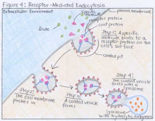
Representation of the plasma membrane   A                                                         B
  • In solution A, there are more sugar molecules on the left than the right - the left is more concentrated
  • The partially permeable membrane only allows water molecules through.
  • The water molecules diffuse through the membrane from the right to the left, down a concentration gradient due to random movements as to reach equilibrium
  • In solution B, the left contains more water molecules, so it is more dilute, and the right contains less water, becoming more concentrated.
Water potential
  • Tendency of water to move out of a solution.
  • Depends on two factors:
    -How much water the solution contains in relation to solutes (concentration)
    -How much pressure is being applied
  • Water potential always moves down a water potential gradient (region of high water potential to a region to low water potential) until water potential is same throughout the system - equilibrium.
  • A solution containing a lot of water (dilute) has a higher water potential than a solution containing a little water (concentrated).

  • Increasing pressure on a solution also increases water potential - increases the tendency for water to move out of it.
  • Water potential of pure water at atmospheric pressure is 0.
  • Therefore a solution (water with solute/solutes dissolved in it) must have a negative water potential - less than 0.
Solute potential and pressure potential
  • Solute potential: Extent to which solute molecules decrease the water potential of the solution.
  • Solute potential is also 0 for pure water, and a negative value for a solution
  • Adding more solute to a solution decreases it's water potential.
  • Pressure potential: Contribution of pressure to the water potential of a solution.
  • Increasing pressure increases water potential.
Osmosis in animal cells
  • If the water potential surrounding the cell is too high - cell swells and bursts (lysis)
  • If it is too low - cell shrinks
  • Essential to maintain a constant water potential in the bodies of animals 
Osmosis in a blood cell

  • Hypotonic: Water potential surrounding cells too high
  • Isotonic: Normal water potential surrounding cells
  • Hypertonic: Water potential surrounding cells too low 
Osmosis in plant cells
  • Unlike animal cells, plants have a cell wall.
  • Cell walls are freely permeable.
  • When water potential is higher outside than inside the cell, water will enter the cell. 
  • However, as water enters plant cells the cell wall will push back against the expanding protoplast (living part of the cell) and pressure builds up.
  • This is the pressure potential and it increases the water potential until the water potential inside and outside the cell are equal - equilibrium is reached
  • The cell wall is so inelastic that it takes very little water to enter the cell to achieve this.
  • Cell wall prevents cell from bursting.
  • A plant cell is fully turgid when it is fully inflated with water.
  • Water potential = Solute potential + Pressure potential 
Osmosis in a plant cell
  • Hypotonic: Fully turgid
  • Isotonic: No net movement of water
  • Hypertonic: Water leaves cell and protoplast gradually shrinks until it exerts no pressure on the cell wall - pressure potential 0.
    As the protoplast continues shrinking it
    begins to pull away from the cell wall.
    This is called
    plasmolysis.
    When the protoplast has completely shrunken away from the cell wall, it is said to be
    fully plasmolysed. When the pressure potential has just reached 0 and plasmolysis is about to occur is called incipient plasmolysis

Active transport 
  • Movement of molecules or ions through carrier proteins across a cell membrane against their concentration gradient using energy from ATP.
  • ATP (produced during cell respiration) is used to make the carrier proteins change its shape, transferring molecules or ions across the membrane in the process.
  • Can occur either into or out of the cell.
  • Example: Sodium potassium (Na+ - K+) pump
  • Found in cell surface membranes of all animal cells
  • Run almost all the time, on average use 30% of a cells energy.
  • For each ATP molecule used it pumps 3 sodium ions out of the cell while at the same time allowing 2 potassium ions into the cell.
  • Net result is the inside of the cell becomes more negative than outside - potential difference (p.d) is created across membrane.
  • The pump has a receptor for ATP on it's inner surface. The receptor acts as an ATPase enzyme in bringing about the hydrolysis of ATP to ADP (adenosine diphosphate) and phosphate to release energy.
Sodium potassium pump

  • Active transport is also important in re-absorption of ions and useful molecules in the kidneys after filtration and absorption of some products of digestion from the gut. In plants, active transport is used inorganic ions from soil to the root hairs and load sugars into the phloem tissues.

Bulk transport

  • Bulk transport of large quantities of materials into (endocytosis) and out of (exocytosis) of cells.
  • Endocytosis: Engulfing of a material by the cell surface membrane to form a small sac (endocytic vacuole). Two forms:
         -
    Phagocytosis (cell eating): Bulk uptake of solid material. Cells specialising in this are called phagocytes, and the vacuoles are called phagocytic vacuoles. Example: White blood cells engulfing bacteria. Phagocytic vacuoles fuse with lysosomes, which contain digestive enzymes.
         -
    Pinocytosis (cell drinking): Bulk uptake of liquid. Vacuoles formed are often really small, in which the process is called micropinocytosis
  • Protein receptors on the outer cell surface membrane detect the molecules that need to be transported and binds to them.
Endocytosis

  • Exocytosis: Reverse of endocytosis - materials are removed from cells. Usually involves golgi body. Example: In secretion of enzymes, secretory vesicles from the golgi body carry the enzymes to the cell surface to release their contents
Exocytosis

 Simplified exocytosis and endocytosis


Thursday, November 26, 2015

Biology: Chapter 4: Cell surface membrane: Cell signalling

Biology: Chapter 4: Cell surface membrane: Cell signalling

  • Getting a message from one place to another.
  • Complex range of signalling pathways which coordinate activities of the cell so they respond appropriately to the environment, even if the cells are large distances apart in the body.
  • Signalling pathway includes:
  1. Receptor receiving a stimulus or signal
  2. Transduction: Converting the signal to a message that is readable 
  3. Transmission of message/signal to effector (target)
  4. The effector making an appropriate response

  • Distances can be short (diffusion within a cell) or long (transport through blood)
    -Endocrine signalling: Signalling over large distances, often through circulatory system
    -Paracrine signalling: Signalling occurring between cells close together either through extracellular fluid or directly between cells
    -Autocrine signalling: Cell stimulates response within itself by releasing signals for it's own receptors

  • Many components and different mechanisms along the route
  • Signalling includes both electrical (nerves) and chemical (hormones).
  • Stimuli can be from inside (hormones) as well as outside (light).
  • The cell surface membrane is a important component of most signalling pathways because it controls what molecules move in and out of the cell.
  • In a typical signalling pathway, molecules must cross or interact with the cell surface membrane.
  • Hydrophobic signalling molecules eg. steroid hormones can diffuse directly through the cell surface membrane and bind to receptors in the cytoplasm or nucleus.
  • Signalling molecules are usually water-soluble.

Typical signalling pathway (for water soluble signalling molecules):

  1. Signal arrives at a protein receptor in the cell surface membrane.
    Receptor is a specific shape which recognizes the signal. Only cells with this receptor can recognize the signal.
  2. Signal changes the shape of the receptor, and since this spans the membrane, the message is in effect passed to the inside of the cell (signal transduction).
    Changing the shape of the reactor allows it to interact with the next component of the pathway, so the message gets transmitted.
  3. The next component is usually a G-protein - a small molecule which diffuses through the cell relaying the message, like a switch to bring about the release of a 'second messenger'.
    G-proteins got their name because the switch mechanism involves binding to GTP molecules - similar to ATP, but with guanine instead of adenine.
  4. After 1 receptor molecule is stimulated, many second messenger molecules can be made in response - amplification, a key feature of signalling.
    Second messenger usually activates an enzyme, which in turn activates further enzymes, increasing amplification at each stage.
    Signalling cascade: Amplification triggered by G-protein.
  5. Response is produced.
Diagram of how cell signalling works.

Other ways receptors alter activity of the cell:

  • Opening an ion channel, resulting in change of membrane potential
  • Acting directly as a membrane-bound enzyme
  • Act as a intracellular (inside the cell) receptor when initial signal passes through