Biology: Chapter 5: Mitotic Cell cycle: Components of a cell cycle
- Cells reproduce by dividing and passing on copies of their genes to 'daughter' cells.
- This is done through nuclear and cell division.
Chromosomes
- Condensed form of chromatin that occurs only during mitosis. Chromatin during normal cell activity (interphase). Consist of 2 identical sister chromatids connected by a centromere.
- Each chromatid contains 1 DNA molecule - chromatids in chromosomes are identical to one another.
 Chromosome during Anaphase
- DNA: Molecule of inheritance. Made up of a series of genes. Only 2nm wide, but total length of the DNA in all the 46 chromosomes in an adult is approximately 1.8 meters. DNA is wound around histones precisely to keep them from being tangled.
- Gene: 1 unit of inheritance. Codes for 1 polypeptide that is involved in a specific aspect of the functioning of the organism.
- Histone: Protein that DNA wounds around. Proteins are basic (alkali) which means they can easily interact with acidic DNA.
- Nucleosome: DNA wrapped around 8 histone molecules. Are like beads on a necklace. Each 'bead' is one nucleosome. Further coiled into chromatin.
- Chromatin: Coiling of DNA around histones (proteins). Makes up chromosomes; chromosomes are the condensed form of chromatin during nuclear division. Easily stained. Exists in two forms: Euchromatin (loosely coiled) and heterochromatin (tightly coiled). During normal cell activity (interphase) chromatin is mostly in the form of Euchromatin so the coding can easily be accessed.
- Double structure - two identical structures called chromatids joined together by a centromere.
 From DNA to chromosomes
- Centromere: Narrow region of the chromosome that joins both chromatids together. Position of the centromere is a particular characteristic for each chromosome. The centromere can be found anywhere along the chromosome. Needed for separation of chromatids during mitosis. Site of attachment for spindle molecules.
- Kinetochore: Attached to centromere during metaphase chromosome, 1 for each chromatid. Made of protein molecules which bind specifically to DNA in the centromere and also to the microtubule spindles. Construction of kinetochore begins before nuclear division (interphase) and are lost after nuclear division.
- Spindle attached to kinetochore of chromosomes pulls the kinetochore, pulling apart the chromosome. This is done by the shortening of the microtubules from both the centrosome pole end and the kinetochore end.
- Centrosomes: Poles of the spindle. Help in the construction of the spindle microtubules, but do not produce the spindle fiber. Centrosomes consist of a pair of centrioles surrounded by large number of proteins that control production of the spindle (the centrioles are not involved in the Microtubule production). Plants do not have centrosomes.
Telomeres
- Telomeres: Made up of multiple DNA base repeat sequences (repeat of guanine (G) for one strand and cytosine (C) for another).
- Have no useful information, but allow enzymes to complete copying important DNA.
- Telomeres are attached to the ends of chromosomes to prevent the genes at the ends of the DNA strand being cut off during replication - if part of the DNA is not copied, the information is lost, which will eventually cause cell death when vital genes are lost.
- Prevents loss of genes during division and allow continued replication.
 Telomeres in cell division
 - Telomerase: Enzyme that adds extra DNA to the telomeres.
- Some cells (mainly specialised cells) do not have telomerase, so with each division their telomeres get a little shorter until vital DNA is no longer protected and the cell dies.
- Could be a mechanism of aging.
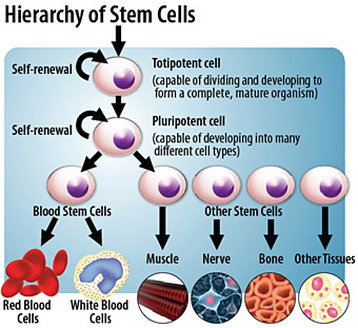
Stem cells
- Cell that can divide an unlimited number of times by mitosis.
- Each time it divides, it has the potential to stay as a stem cell or develop into a specialized cell (eg. nerve cell).
- Potency: How many different cell types the stem cell can specialize into.
- Totipotent: Stem cells that can produce any type of cell. Zygotes (fertilization of sperm and egg) and cells up to 16-stage development are totipotent,
- Pluripotent: Embryonic stem cells after some cells have specialized to form the placenta. The cells can no longer form a placenta, but can form every other cell that leads to the development of the embryo and later the adult.
- Multipotent: Stem cells that are only able to form a few types of cells eg. bone marrow cells can form red and white blood cells. Can replicate any number of times but can only produce those type of cells.
- The more cells specialize, the more they lose the ability to divide. In an adult most cells do not divide.
- Stem cell therapy: Introduction of new adult stem cells into damaged tissue to treat disease or injury. Bone marrow transplantation is the only form of this therapy that has progressed passed experimental stage into routine medical practice. Experiments with growing new tissues or even organs from isolated stem cells in laboratories have been conducted.
Cancer
- Result of uncontrolled mitosis.
- Divide repeatedly and form a tumor
- Tumor: Irregular mass of cells, which usually show abnormal changes in shape. Typical tumor contains approximate 1000 million cancerous cells.
- Thought to start when changes occur in the gene that cause cell division.
- Mutation: Change in any gene.
- Oncogene: Mutated gene that causes cancer. 'Onkos' means bulk or mass in greek.
- Mutations occur frequently and don't usually lead to cancer, usually getting destroyed by the immune system or getting affected in a way that leads to an early death, and get replaced by mitosis.
- Cancerous cells manage to bypass cell checkpoints in the interphase and cell death, leading to the cancerous cell replicating and passing on faulty genes to all of it's descendants.
- Carcinogen: Factor that causes cancer eg. pollution, radiation, UV light
- Not all tumors are cancerous
- Benign tumors: Non-cancerous tumors - do not spread from site of origin eg. warts
- Malignant tumors: Spread through the body, invading other tissues and destroying them. Interfere with the normal functioning of the area where they started to grow; may block off intestines, lungs or blood vessels.
- Metastasis: Cells that break off and spread through the blood and lymphatic system (transport lymph) and form secondary growths.
Most dangerous characteristic of cancer.
Can be hard to find and remove secondary cancers.
 Normal cell vs cancer cell
No comments:
Post a Comment